

Product datasheet
FITC Annexin V Apoptosis Detection Kit with PI
Product Information
Catalog / Size PDZK-01 / 100 tests
Storage Temperature 2–8 °C. Do not freeze.
Kit Components 10X Annexin V Binding Buffer (10 mL)
- Contains aqueous buffered solution containing 0.1 M Hepes/NaOH (pH 7.4), 1.4 M NaCl, 25 mM CaCl2 with no preservative.
- Prepare Freshly. For a 1X working solution, dilute 1 part of the 10X Annexin V Binding Buffer to 9 parts of distilled water.
Annexin V FITC (100 µL)
- Volume recommends per test; 1 µL
- Contains aqueous buffered solution containing BSA and ≤0.09% sodium azide.
Propidium Iodide (PI) Staining Solution (100 µL)
- Volume recommends per test; 1 µL
- Contains aqueous buffered solution containing no preservative.
The components supplied in this kit are 0.2 µm filtered and aseptically filled.
Storage Note Store undiluted at 4°C and protect from prolonged exposure to light. Do not freeze.
Reactivity All mammalian species
Application Flow Cytometry – Quality Tested
Kit Properties
FITC Annexin V Apoptosis Detection Kit with PI has been specifically designed for the identification of apoptotic and necrotic cells. Annexin V FITC serves as a fluorescent probe for apoptotic cells. They will not bind normal and intact cells. However, necrotic cells are leaky enough to pass Annexin V FITC into cytoplasm and provide accessibility for binding to inner membrane phosphatidylserine (false positive apoptosis). Indeed, apoptotic cells have to be differentiated from necrotic cells by PI-stained apoptotic nuclei in comparison to PI-stained necrotic nuclei. But this assay due to its distinctive design is probably able to distinguish between cells that have undergone apoptotic death versus those that have died as a result of a necrotic pathway. The dead cells will probably stain only with both PI. However, when apoptosis is measured over time, cells can be often tracked from FITC Annexin V and PI negative (viable, or no measurable apoptosis), to FITC Annexin V positive and PI negative (early apoptosis, membrane integrity is yet present) and finally to FITC Annexin V and PI positive (end stage apoptosis). In fact, the movement of cells through these three stages suggests apoptosis (Figs. 1-3)
Description
Apoptosis is a normal physiologic process which occurs during embryonic development as well as in maintenance of tissue homeostasis. However, the program cell death is triggered in non-physiological conditions such as, drug induced apoptosis. The apoptosis program is characterized by certain morphological features, including loss of plasma membrane asymmetry at earlier stage and at later stage distinguish with subsequent loss of membrane integrity, condensation of the cytoplasm and nucleus, and internucleosomal cleavage of DNA. In apoptotic cells, the membrane phospholipid phosphatidylserine (PS) is translocated from the inner to the outer leaflet of the plasma membrane, thereby PS exposes to the extracellular environment. Annexin V is a 35-36 kDa Ca2+ dependent phospholipid-binding protein that binds phosphatidylserine with high affinity.
The conjugation of Annexin V to fluorochromes including FITC, while retains its high affinity for PS, provide this conjugate as a sensitive probe for flow cytometric analysis of cells that are undergoing apoptosis and microscopic imaging of this stained population. Since externalization of PS occurs in the earlier stages of apoptosis, FITC Annexin V staining can identify apoptosis at an earlier stage, and then quantitative evaluation of apoptotic nuclei based on nuclear changes such as DNA fragmentation can be distinguished by fluorophore staining nuclei, such as propidium iodide (PI) or 7-Amino-Actinomycin (7-AAD) to allow the investigator to identify early apoptotic cells (PI or 7-AAD negative, FITC Annexin V positive).

Figure 1: Test principle.
Viable and dead cells Viable cells with intact membranes represent preserved plasma membrane integrity so are not permeable to PI, whereas the membranes of dead and damaged cells are permeable to PI. For example, cells that are considered viable are FITC Annexin V and PI negative
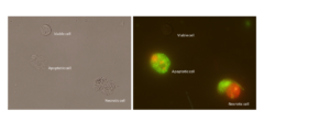
Figure 2: The illustration of viable, necrotic and apoptotic cells.
Early apoptotic cells The cells in early apoptosis population are FITC Annexin V positive and PI negative
Late apoptotic cells The cells in late apoptosis or already dead are both FITC Annexin V and PI positive.

Figure 3: The illustration of viable, early and late apoptotic cells.
Flow Cytometric Analysis of FITC Annexin V/PI staining
| Jurkat cells (Human T-cell leukemia; ATCC TIB-152) were untreated on left panels
or treated on right panels during three time points, 3, 6 and 18 hours with 1 μM staurosporine (right panels). Cells were incubated with FITC Annexin V in binding buffer and then incubated with propidium iodide (PI) and analyzed by flow cytometry. At left panels, untreated cells at three time points were FITC Annexin V and PI negative, indicating that they were viable and not undergoing apoptosis. At right panels, after 3 hours treatment (top panel), after 6 hours treatment (middle panel) and after 18 hour treatment (bottom panel) there were two populations of cells: Cells that were viable and not undergoing apoptosis (FITC Annexin V and PI negative; Q4 area in each panel) and cells undergoing early apoptosis (FITC Annexin V positive and PI negative; Q3 area) and late apoptosis (FITC Annexin V positive and PI positive; Q2 area) (Fig. 4).
Figure 4. Flow Cytometric Analysis of FITC Annexin V staining. |

Materials
– Jurkat T cells (ATCC TIB-152).
– Prepare staurosporine (abcam; ab120056): final concentration 1µM of staurosporine in cell culture media contains 1 x 10⁵ – 1 x 10⁶ cells/mL.
– 10X Annexin V Binding Buffer (component no. PZ03071): Prepare 1X working Solution through dilution 1 part of the 10X Annexin V Binding Buffer to 9 parts of distilled water.
– FITC Annexin V (component no. PDZ233): Use 1 μl per test.
– Propidium Iodide (PI) (component no. PZ03072) is ready-to-use as a nucleic acid dye. Use 1 μl per test.
Staining Procedure
1- Wash cells with 1X Binding Buffer through centrifugation at 300g, 7 min, without breaking.
2- Resuspend cells in 1X Binding Buffer at a concentration of 1 x 10⁵ to 1 x 10⁶ cells in 100µL.
3– Add 1 μl of Annexin V FITC to the test tube.
4– Gently vortex the cells and incubate for 15 min at RT (25°C) in the dark.
5– Add 1 μl of PI to the test tube and incubate for 5 minutes.
6– Wash the tubes with 1X PBS through centrifugation at 300g, 10 min (without breaking).
7– Add 400 μl of 1X PBS to each tube. Analyze by flow cytometry within 1 hr, while samples keep on ice.
Required Controls for Setting up flow cytometry
| – Unstained cells to set up dot plot quadrant.
– Cells stained with Annexin V FITC (no PI), as a single FITC color population while has to contain negative cells to set up compensation. – Cells stained with PI (no FITC Annexin V), as a single PI color population while has to contain negative cells to set up compensation. – Untreated normal cells incubated with Annexin V FITC and PI that determines the baseline of apoptosis in normal cell population and is described as auto-apoptosis. |
References
1- Simonian M, Haji Ghaffari M, Salimi A, Mirzadegan E, Sadeghi N, Ebrahimnezhad N, Fazli G, Fatemi R, Bayat AA, Milani S, Negahdari B, Rabbani H. Monoclonal Antibody Against Sortilin Induces Apoptosis in Human Breast Cancer Cells. Avicenna J Med Biotechnol. 2022 Jan-Mar;14(1):37-45. doi: 10.18502/ajmb.v14i1.8168. PMID: 35509360; PMCID: PMC9017463.
2- Sohbati H, Amini M, Balalaie S. Synthesis and Biological Evaluation of Novel Anti-leukemia Proteolysis-Targeting Chimeras in Degradating Inosine Monophosphate Dehydrogenase. Iranian Journal of Pharmaceutical Research. 2022 Dec 31;21(1).
3- Ghaffari MH, Salimi A, Mirzadegan E, Sadeghi N, Ebrahimnezhad N, Fazli G, Fatemi R, Bayat AA, Negahdari B, Rabbani H. Monoclonal Antibody Against Sortilin Induces Apoptosis in Human Breast Cancer Cells. researchsquare.com. 2021
4- Moradipour A, Dariushnejad H, Ahmadizadeh C, Lashgarian HE. Dietary flavonoid carvacrol triggers the apoptosis of human breast cancer MCF-7 cells via the p53/Bax/Bcl-2 axis. Medical Oncology. 2022 Dec 10;40(1):46.
Please note: All products are “FOR RESEARCH USE ONLY. NOT FOR USE IN DIAGNOSTIC PROCEDURES”


نقد و بررسیها
هنوز بررسیای ثبت نشده است.