

Product Datasheet
Mouse Anti-Human beta-actin
Overview
Product number PDZMM114
Host species Mouse
Target species Human, Horse, Rat, Dog, Bovine, Monkey
Suitable for: IHC-P, WB, ELISA, Immunomicroscopy, Dot blot, ICC, IHC-Fr
Immunogen A KLH-conjugated synthetic peptide derived from N-terminal region of Beta-actin protein was used for immunization.
Conjugation Unconjugated
Properties
Form Liquid
Storage instructions Shipped at 4 °C. Store at -20 °C. Avoid freeze/thaw cycle. Please see notes section.
Storage buffer Phosphate buffered saline pH 7.4, contains stabilizer and ≤0.09% sodium azide.
Purity Immunogen affinity or SpG purified
Purification notes This product was prepared by immunoaffinity chromatography using immunogen peptide coupled to Sepharose 4B.
Conjugation notes –
Clonality Monoclonal
Isotype IgG
General notes For extended storage aliquot contents and freeze at -20 °C or below. Centrifuge product if not completely clear after standing at room temperature. This product is stable for several weeks at 4 °C as an undiluted liquid. Dilute only prior to immediate use.
Our customer’s feedback says the antibody worked great. If in case the antibody fails to give results then please contact our scientific support team for assistance.
Applications
The application notes include recommended starting dilutions; optimal dilutions/concentrations should be determined by the end-user.
Product Usage Information:
Application Dilutions
Western Blotting 3-5 ug/ml
Immunohistochemistry (Paraffin) 5-10 ug/ml
Immunohistochemistry (Frozen) 5-10 ug/ml
Immunofluorescence 5-10 ug/ml
Flow Cytometry 5-10 ug/ml
Please note: All products are “FOR RESEARCH USE ONLY. NOT FOR USE IN DIAGNOSTIC PROCEDURES”
Background:
Actin, a ubiquitous eukaryotic protein, is the major component of the cytoskeleton. At least six isoforms are known in mammals. Nonmuscle β- and γ-actin, also known as cytoplasmic actin, are predominantly expressed in nonmuscle cells, controlling cell structure and motility (1). α-cardiac and α-skeletal actin are expressed in striated cardiac and skeletal muscles, respectively; two smooth muscle actins, α- and γ-actin, are found primarily in vascular smooth muscle and enteric smooth muscle, respectively. These actin isoforms regulate the contractile potential of muscle cells (1). Actin exists mainly as a fibrous polymer, F-actin. In response to cytoskeletal reorganizing signals during processes such as cytokinesis, endocytosis, or stress, cofilin promotes fragmentation and depolymerization of F-actin, resulting in an increase in the monomeric globular form, G-actin (2). The ARP2/3 complex stabilizes F-actin fragments and promotes formation of new actin filaments (2). Research studies have shown that actin is hyperphosphorylated in primary breast tumors (3). Cleavage of actin under apoptotic conditions has been observed in vitro and in cardiac and skeletal muscle, as shown in research studies (4-6). Actin cleavage by caspase-3 may accelerate ubiquitin/proteasome-dependent muscle proteolysis (6).
Beta actin is one of six actin isoforms which have been identified. Beta actin is a non-muscle cytoskeletal protein in all human cell types and is involved in cell motility, structure, and integrity. There are six different actin isoforms in human. The beta isoform of actin, along with gamma actin, coexist in most cell types as components of the cyto-skeleton. Beta actins are cytoplasmic proteins ubiquitously expressed in all eukaryotic cells. Polymerization of globular actin (G-actin) leads to a structural filament (F-actin) in the form of two stranded helix. Actins are highly conserved proteins that are involved in cell motility, structure and integrity. Because beta actin is ubiquitously expressed in all eukaryotic cells, it is frequently used as a loading control for assays involving protein detection, such as Western blotting. Antibodies to beta-actin provide a specific and useful tool in studying the intracellular distribution of beta-actin and the static and dynamic aspects of the cytoskeleton.
Storage:
Supplied in 10 mM sodium HEPES (pH 7.5), 150 mM NaCl, 100 µg/ml BSA and 50% glycerol. Store at –20°C. After receiving aliquot the antibody and store at –20°C.
Terms and conditions
Guarantee only valid for products bought direct from PADZA or one of our authorized distributors
References:
- Herman, I.M. (1993) Curr Opin Cell Biol 5, 48-55.
- Condeelis, J. (2001) Trends Cell Biol 11, 288-93.
- Lim, Y.P. et al. (2004) Clin. Cancer Res. 10, 3980-3987.
- Kayalar, C. et al. (1996) Proc. Natl. Acad. Sci. USA. 93, 2234-2238.
- Communal, C. et al. (2002) Proc. Natl. Acad. Sci. USA. 99, 6252-6256.
- Du, J. et al. (2004) J. Clin. Invest. 113, 115-123.
The product has been utilized in:
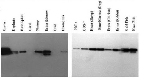
Analysis of reactivity of anti-β-actin antibody with lysates from a panel of organisms with different origins by western blot. A panel of tissue samples from different organisms were lysed and subjected to western blot under reducing condition.
Note: This product has originally been developed at Avicenna Research Institute, Tehran, IRAN and assigned to PADZA Company according to contract 98/15/191dated 98/01/10.

نقد و بررسیها
هنوز بررسیای ثبت نشده است.